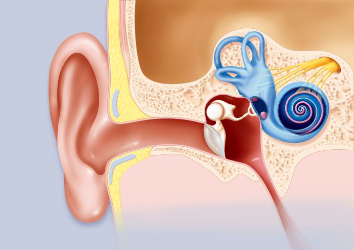
Label the structures of the cochlea and embark on a captivating journey into the intricate world of hearing. This comprehensive guide delves into the anatomy, physiology, and clinical significance of the cochlea, providing a thorough understanding of this remarkable organ’s role in sound perception.
The cochlea, a spiral-shaped structure nestled within the inner ear, plays a pivotal role in converting sound waves into electrical signals that the brain interprets as sound. Its intricate internal architecture, comprising the scala tympani, scala vestibuli, and scala media, enables the cochlea to perform this remarkable feat.
External Anatomy of the Cochlea
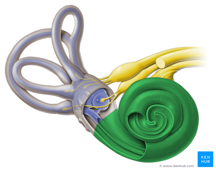
The cochlea, a crucial component of the inner ear, is a small, snail-shaped structure located deep within the temporal bone. Its primary function is to convert sound waves into electrical signals that can be interpreted by the brain. The cochlea is composed of both bony and cartilaginous components.The
bony portion of the cochlea, known as the osseous labyrinth, is a complex network of canals and chambers that form the external structure of the cochlea. It is made up of three main parts: the scala vestibuli, scala tympani, and scala media.
The scala vestibuli is the upper chamber, while the scala tympani is the lower chamber. The scala media, located between the other two chambers, is filled with a fluid called endolymph.The cartilaginous portion of the cochlea, known as the membranous labyrinth, is a delicate, tube-like structure suspended within the osseous labyrinth.
It is composed of the vestibular membrane, basilar membrane, and tectorial membrane. These membranes play a crucial role in the transmission of sound waves and the generation of electrical signals.Together, the bony and cartilaginous components of the cochlea form a protective and supportive environment for the delicate structures responsible for hearing.
The unique shape and composition of the cochlea enable it to amplify and filter sound waves, allowing us to perceive a wide range of sounds and frequencies.
Internal Anatomy of the Cochlea
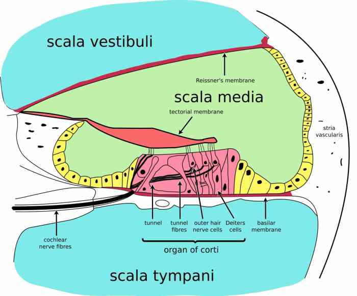
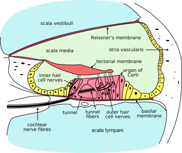
The cochlea is a spiral-shaped structure in the inner ear responsible for hearing. Its internal anatomy consists of three fluid-filled chambers called scalae: scala tympani, scala vestibuli, and scala media.
Scala Tympani, Scala Vestibuli, and Scala Media, Label the structures of the cochlea
The scala tympani and scala vestibuli are filled with perilymph, a fluid similar to cerebrospinal fluid. The scala media is filled with endolymph, a fluid with a different ionic composition than perilymph.
The scala tympani and scala vestibuli are separated by the basilar membrane, a thin, flexible membrane that plays a crucial role in sound perception.
Basilar Membrane
The basilar membrane is wider and thicker at the base of the cochlea and narrower and thinner at the apex. When sound waves enter the cochlea, they cause the basilar membrane to vibrate. The frequency of the sound wave determines the location of maximum vibration along the basilar membrane.
Low-frequency sounds cause vibrations near the base of the cochlea, while high-frequency sounds cause vibrations near the apex.
Organ of Corti
The organ of Corti is a sensory structure located on the basilar membrane. It contains specialized sensory cells called hair cells, which are responsible for converting sound vibrations into electrical signals.
The hair cells are arranged in rows, with inner hair cells and outer hair cells. Inner hair cells are responsible for transmitting sound information to the brain, while outer hair cells amplify and fine-tune the sound signals.
Physiology of Hearing
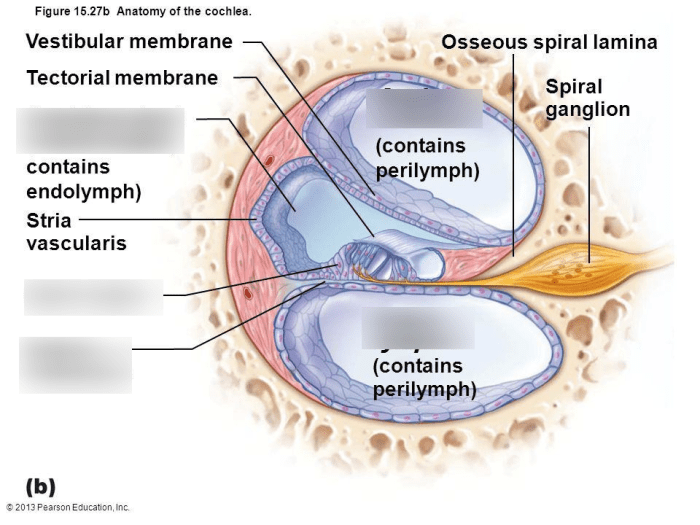
The physiology of hearing involves the intricate process of sound wave transmission and transduction within the cochlea, leading to the perception of sound.
Sound Wave Transmission through the Cochlea
Sound waves, upon reaching the eardrum, set it into vibrations. These vibrations are transmitted through the ossicles (malleus, incus, and stapes) to the oval window, which in turn transmits them to the fluid within the cochlea. The sound waves then travel through the perilymph in the scala vestibuli and scala tympani, causing the basilar membrane to vibrate.
Role of the Tectorial Membrane in Auditory Transduction
The tectorial membrane is a gelatinous structure that overlies the basilar membrane. When the basilar membrane vibrates, the tectorial membrane moves in the opposite direction, causing shear forces between the two membranes. These shear forces stimulate the hair cells located on the basilar membrane, initiating the process of auditory transduction.
Tonotopic Organization of the Cochlea
The cochlea exhibits a tonotopic organization, meaning different frequencies of sound are detected at specific locations along its length. High-frequency sounds are detected near the base of the cochlea, while low-frequency sounds are detected near the apex. This tonotopic organization allows for the accurate localization and discrimination of sound frequencies.
Clinical Significance of Cochlear Structures
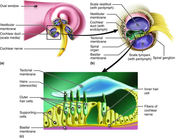
The intricate structures of the cochlea play a crucial role in our ability to hear and process sound. Understanding the clinical significance of these structures is paramount for diagnosing and treating hearing impairments.
Cochlear Malformations and Disorders
Cochlear malformations and disorders can result from genetic abnormalities, environmental factors, or infections. These conditions can affect the shape, size, or function of the cochlea, leading to varying degrees of hearing loss.
- Mondini malformation:A congenital anomaly characterized by a shortened and malformed cochlea, resulting in profound hearing loss.
- Scheibe deformity:A cochlear malformation where the cochlear duct is narrow and partially or completely divided, causing conductive or sensorineural hearing loss.
- Cochlear otosclerosis:A condition in which abnormal bone growth in the cochlea fixes the stapes footplate, resulting in conductive hearing loss.
- Ménière’s disease:A disorder of the inner ear characterized by episodes of vertigo, hearing loss, and tinnitus, potentially caused by fluid buildup in the cochlea.
Cochlear Implants
Cochlear implants are electronic devices that provide a sense of hearing to individuals with severe to profound hearing loss. They work by bypassing the damaged cochlea and directly stimulating the auditory nerve.
- Indications:Cochlear implants are recommended for individuals with severe to profound hearing loss who do not benefit from hearing aids.
- Procedure:The implant is surgically placed into the cochlea, and electrodes are inserted into the cochlear duct to stimulate the auditory nerve.
- Outcomes:Cochlear implants can significantly improve hearing and speech understanding, allowing individuals to participate more fully in daily life.
Importance of Understanding Cochlear Structures
Understanding the cochlea’s anatomy and physiology is essential for:
- Accurate diagnosis:Identifying the underlying cause of hearing loss by assessing the integrity of cochlear structures.
- Effective treatment:Developing appropriate treatment plans, such as cochlear implants, based on the specific cochlear abnormality.
- Prevention:Implementing preventive measures to minimize the risk of cochlear damage and hearing loss.
Common Queries: Label The Structures Of The Cochlea
What is the function of the basilar membrane in the cochlea?
The basilar membrane plays a crucial role in sound perception by vibrating in response to sound waves. Its stiffness and width vary along its length, allowing it to resonate at different frequencies, enabling the cochlea to distinguish between different pitches.
What are the sensory cells located in the organ of Corti?
The sensory cells in the organ of Corti are hair cells, which are specialized cells that convert mechanical vibrations into electrical signals. These signals are then transmitted to the brain via the auditory nerve.
How do cochlear implants work?
Cochlear implants are surgically implanted devices that bypass damaged hair cells in the cochlea and directly stimulate the auditory nerve. This allows individuals with severe hearing loss to regain some degree of hearing.
